Non-boring, neither realistic SKIN DIAGRAMS
About it
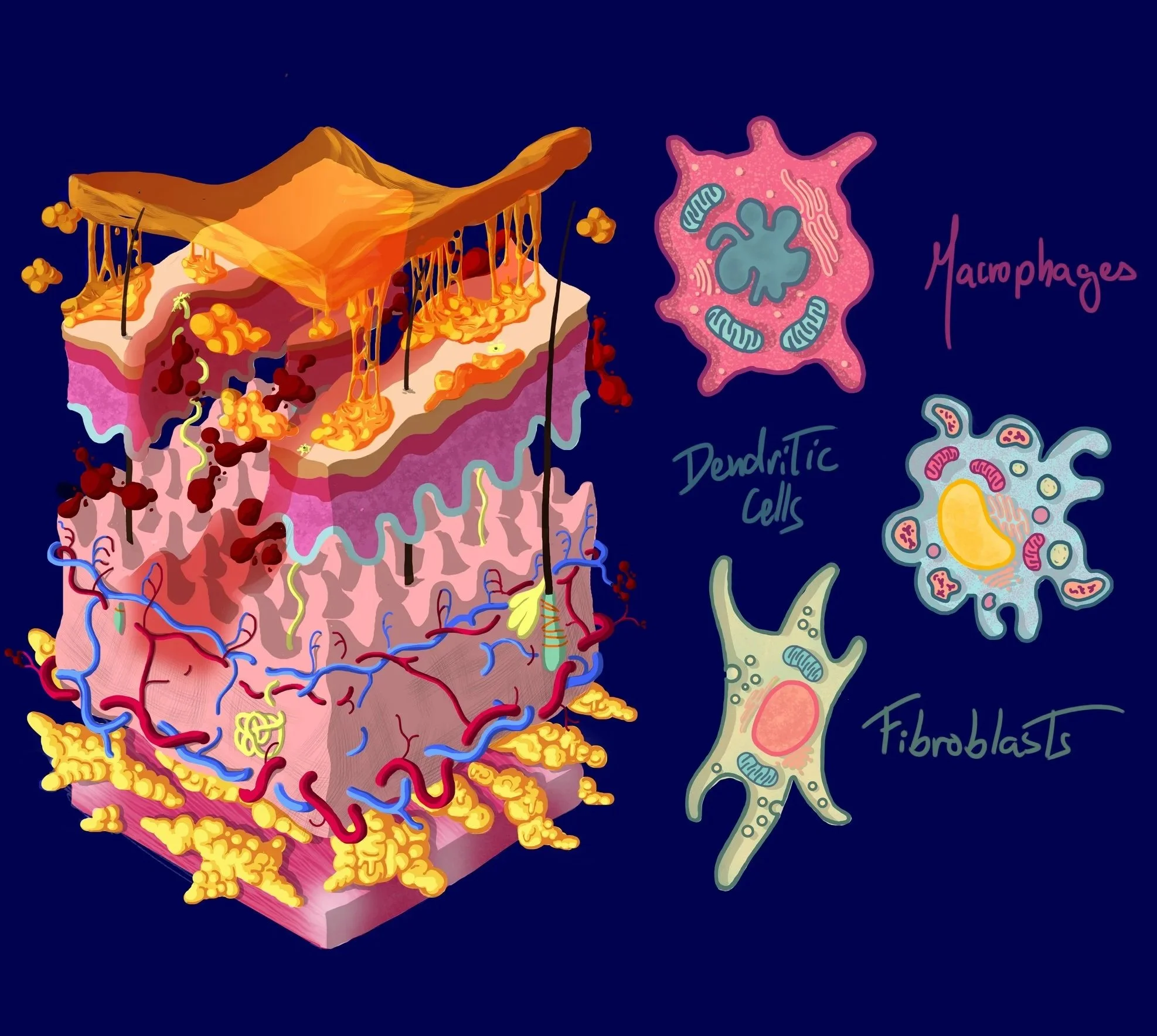
This illustration was conceived as the TOC graphic for a wound-healing deep eutectic solvent paper by Dr Sergio Martin Saldaña. The materials at the centre of the study interact with cells like fibroblasts and macrophages, modulating their response and accelerating the healing of skin lesions, opening new possibilities in regenerative medicine.
Roles: Conceptualisation, Illustration
Client: POLYMAT & ICTP-CSIC
Tools: Photoshop
The Challenge
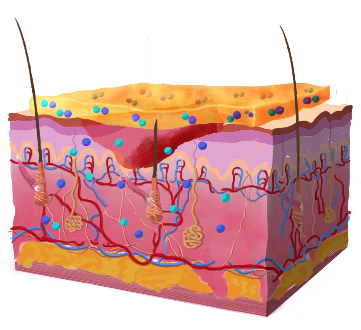
Skin diagrams are everywhere in biomedical literature, and they almost always look the same: flat, technical, lifeless outlines. The challenge was to represent a traditional skin cross-section in a way that felt alive and visually striking, while still carrying the scientific content from the paper, such as the layered architecture, the key differential cells, and the role of adhesive gel-based materials for tissue regeneration.
The Process
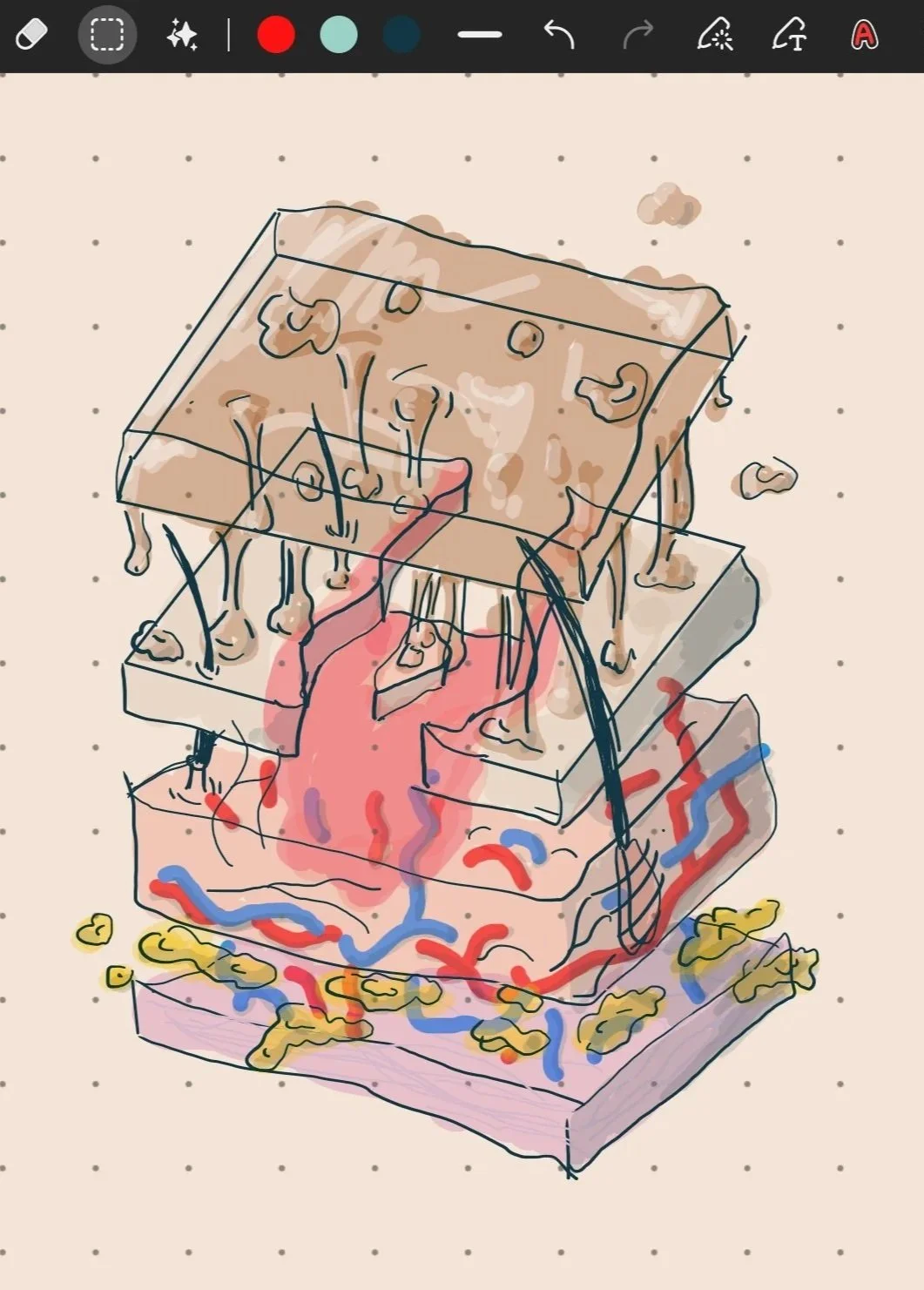
I built the skin as an isometric model with an impressionistic, anti-gravitational quality, full of textures designed to grab attention. Each layer becomes a colourful, almost abstract landscape rather than a labelled slice, making even the most technical details something you want to look at before you read about them.
The key cells of the inflammatory system (macrophages, dendritic cells, and fibroblasts) were redrawn from scratch. Here they aren't mere dots on a graph but living elements packed with organelles, evoking pools filled with floats and joy.
The gel materials were integrated into the scene as tangible, sculptural forms rather than abstract blobs, reinforcing their physical role in modulating the cellular environment and promoting healing.
The whole piece is an attempt to break with the established and explore something less expected, to show that art, science, and creativity can coexist and offer a new way of looking at what has always been in front of us.